 English
 ไทย
 မြန်မာ
 中文
 العربية
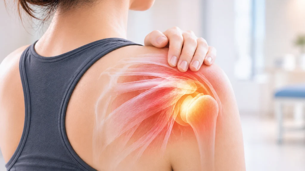
Individuals who begin experiencing shoulder pain, restricted elevation, or difficulty reaching behind their back often share common questions, such as:
- Is nighttime shoulder pain a sign of frozen shoulder?
- What causes the inability to raise the arm?
- Can frozen shoulder truly resolve on its own?
- How long does frozen shoulder recovery take?
- Is surgery necessary for frozen shoulder?
These inquiries are frequently encountered by clinicians managing shoulder pathology, and the clinical answers are often more complex than anticipated. Frozen shoulder is not merely simple “shoulder joint stiffness”; rather, it is a complex pathological condition characterized by capsular inflammation, contracture, and fibrosis. In many patients, it is also closely linked to systemic metabolic conditions such as diabetes mellitus, thyroid disorders, hyperlipidemia, and metabolic syndrome.
Q&A: What is Frozen Shoulder?
Frozen Shoulder, clinically termed Adhesive Capsulitis, is a pathological state in which the shoulder joint capsule undergoes chronic inflammation, progressive thickening, and contracture, leading to marked loss of joint mobility. Patients typically present with progressive pain accompanied by worsening joint rigidity that substantially disrupts activities of daily living (ADLs).
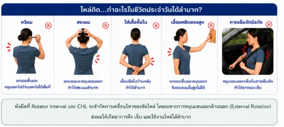
Routine functional tasks that were once effortless—such as combing hair, washing hair, fastening undergarments, fastening a seatbelt, or reaching for overhead objects—become notably difficult and painful.
Q&A: How Common is Frozen Shoulder?
Frozen shoulder is a relatively prevalent musculoskeletal condition, affecting approximately 2–5% of the general population. It predominantly manifests between 40 and 60 years of age, with a slightly higher prevalence observed in females compared to males.
Although the condition frequently arises idiopathically without an identifiable antecedent cause, several patient cohorts exhibit a substantially heightened risk, including:
- Patients diagnosed with diabetes mellitus
- Patients with thyroid disease
- Individuals with hyperlipidemia
- Individuals presenting with visceral obesity or metabolic syndrome
- Patients with a history of shoulder trauma or surgical intervention
Clinical Note: In diabetic populations specifically, the incidence of frozen shoulder is multifold higher than that of non-diabetic cohorts. Furthermore, these patients typically endure more severe symptomatic contractures, prolonged recovery periods, and elevated rates of bilateral involvement or recurrence.
Q&A: What are the Clinical Symptoms of Frozen Shoulder?
Common clinical signs and symptoms include:
- Insidious, non-traumatic shoulder pain
- Exacerbation of pain during nighttime hours
- Inability to lie or sleep on the affected shoulder
- Progressive joint tightness and end-range restriction
- Impaired ability to comb or shampoo hair
- Marked restriction in reaching behind the back (internal rotation)
- Difficulty fastening undergarments or securing a seatbelt
Hallmark Early Clinical Sign: A pronounced, selective loss of passive and active External Rotation. Patients often first notice severe difficulty when washing hair or placing hands behind the head, even when anterior forward flexion remains relatively preserved.
Previous Paradigms Regarding Frozen Shoulder
Historically, the conventional orthopedic belief was that frozen shoulder represented a localized inflammatory and contractive pathology of the glenohumeral capsule culminating in periatricular fibrosis, which would eventually resolve benignly on its own given sufficient time. Consequently, many patients were counseled to simply “wait it out” with standard physical therapy, expecting spontaneous complete resolution.
However, modern translational research has demonstrated that Adhesive Capsulitis is not merely an isolated joint pathology. Rather, it is a complex systemic fibrotic cascade driven by chronic neurovascular inflammation, aberrant matrix remodeling, and systemic metabolic dysregulation (closely correlated with diabetes mellitus, thyroidopathies, dyslipidemia, and metabolic syndrome).
This contemporary paradigm shift has transformed clinical management. Therapeutic regimens no longer focus exclusively on passive symptom suppression or aggressive stretching. Instead, modern management prioritizes accurate clinical staging, early targeted anti-inflammatory intervention, tailored mechanotherapy, and concurrent optimization of systemic metabolic risk factors.
Q&A: Where Does Capsular Fibrosis Predominate? Why is External Rotation Specifically Blocked?
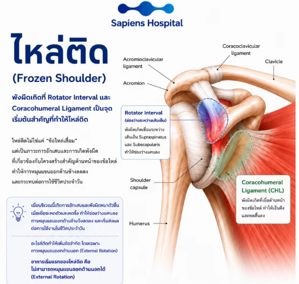
Anatomical, histological, and arthroscopic studies confirm that capsular fibrosis is not uniformly distributed throughout the joint. Instead, pathological tissue thickening and fibrotic contracture are heavily concentrated within the anterior-superior structures of the shoulder joint:
- Rotator Interval: The anatomical interval between the anterior border of the supraspinatus tendon and the superior border of the subscapularis tendon becomes densely fibrotic, leading to obliteration and narrowing of the interval space.
- Coracohumeral Ligament (CHL): This crucial anterior stabilizing ligament undergoes severe fibrotic hyperplasia, becoming markedly thickened, rigid, and contracted.
These two anterior structures serve as the primary anatomical restraints controlling glenohumeral external rotation. When they become chronologically inflamed, thickened, and contracted, external rotation is mechanically blocked before restrictions in other planes of motion become apparent.
How Does This Mechanistic Insight Impact Surgical and Interventional Strategy?
While historic approaches frequently involved non-selective 360-degree pan-capsular release, modern arthroscopic techniques increasingly favor Targeted Capsular Release. By selectively releasing the contracted rotator interval and coracohumeral ligament (CHL), clinicians restore external rotation and functional kinematics with precision while minimizing collateral capsular and neurovascular trauma.
Q&A: Does Glycemic Control Improve Frozen Shoulder? Considerations for Corticosteroid Injections in Diabetic Patients
- Impact of Hyperglycemia: Sustained elevated blood glucose promotes advanced glycation end-products (AGEs) cross-linking, which severely diminishes tensile elasticity in collagen fibers within tendons and the joint capsule, precipitating rapid fibrotic contracture. Poorly controlled diabetic patients consistently present with denser capsular contracture and prolonged recalcitrance. Rigorous glycemic optimization helps suppress systemic low-grade inflammation and fosters microvascular and tissue healing.
- Intra-articular Corticosteroid Injections: Intra-articular corticosteroids provide potent, rapid anti-inflammatory and analgesic relief, substantially expanding the therapeutic window for rehabilitation. However, in diabetic patients, intra-articular steroid absorption frequently induces transient systemic hyperglycemia lasting approximately 2–5 days. Clinicians must advise rigorous blood glucose self-monitoring during this post-injection window.
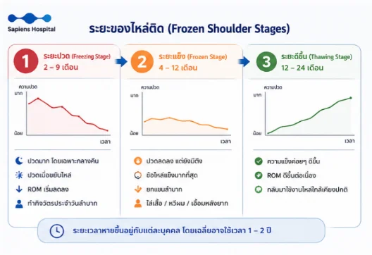
Q&A: Does Frozen Shoulder Truly Self-Resolve? Understanding the Three Clinical Stages of Adhesive Capsulitis
The long-standing clinical dictum that adhesive capsulitis is entirely self-limiting within 1–2 years does not hold true universally. Long-term outcome registries show that a substantial subset of patients continue to suffer from objective range-of-motion (ROM) deficits and residual functional impairment years after symptomatic onset.
The Three Clinical Stages of Frozen Shoulder:
- Stage 1: Freezing Stage (Pain Phase) [Duration: ~2–9 months]:
Characterized by insidious onset of severe, diffuse shoulder pain, notably prominent at night, with marked intolerance to sleeping on the involved side. Range of motion progressively declines as inflammatory synovitis worsens (pain predominates over mechanical contracture). - Stage 2: Frozen Stage (Stiffening Phase) [Duration: ~4–12 months]:
Resting pain gradually attenuates, but capsular fibrosis consolidates into profound mechanical rigidity. Severe restrictions in elevation, external rotation, and internal rotation severely impair activities of daily living (“decreased resting pain, but severe functional joint blockage”). - Stage 3: Thawing Stage (Recovery Phase) [Duration: ~12–24 months]:
Capsular stiffness progressively subsides, with gradual restoration of glenohumeral ROM and return toward baseline functional capacity (although mild terminal ROM deficits may persist permanently in select individuals).
The duration of natural recovery varies widely across individual profiles, averaging 1–2 years. Initiating structured medical intervention early during the inflammatory stage significantly mitigates pain, curtails capsular contracture progression, and prevents entrenched fibrosis.
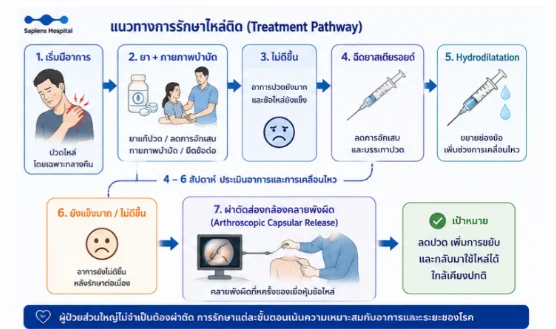
Structured Treatment Pathway for Frozen Shoulder
1. Symptom Onset (Shoulder pain / Progressive loss of ROM)
↓
2. Pharmacologic Analgesia/Anti-inflammatories + Stage-Matched Physical Therapy
↓ (In cases with inadequate improvement)
3. Ultrasound-Guided Intra-Articular Corticosteroid Injection
↓ (Or combined interventional modality)
4. Glenohumeral Hydrodilatation (Capsular Distension)
↓ (For refractory severe stiffness / failed conservative care)
5. Arthroscopic Capsular Release (ACR)
(Primary Objective: Alleviate pain, expand functional range of motion, and restore normal joint biomechanics, with the vast majority of patients successfully managed without requiring surgical intervention).
1. Pain Optimization and Physical Rehabilitation
- Pain Management: Initiated via evidence-based analgesics, NSAIDs, localized cryotherapy/thermotherapy, and activity modification to establish an adequate analgesic window for therapeutic movement.
- Physical Therapy: Remains the cornerstone of functional recovery (aimed at pain reduction, capsular mobilization, and contracture prevention). Crucially, aggressive stretching beyond pain thresholds must be avoided during the acute inflammatory freezing stage, as forced end-range mobilization exacerbates synoviocyte irritation and accelerates capsular reactive fibrosis.
2. Corticosteroid Injections and Hydrodilatation
- Corticosteroid Injection: Targeted intra-articular glenohumeral injection (optimally performed under real-time ultrasound guidance) effectively arrests acute synovial inflammation and provides rapid pain reduction, thereby enabling high-compliance physical rehabilitation.
- Hydrodilatation (Capsular Distension): An interventional procedure involving the controlled high-pressure injection of sterile saline combined with local anesthetic and corticosteroid into the glenohumeral joint space to mechanically distend the contracted capsule, attenuate stiffness, expand capsular volume, and alleviate pain.
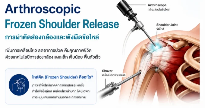
3. Arthroscopic Capsular Release (ACR)
Indicated when comprehensive conservative and interventional management fails to yield clinical progress and persistent, severe capsular restriction severely impairs occupational performance or overall quality of life.
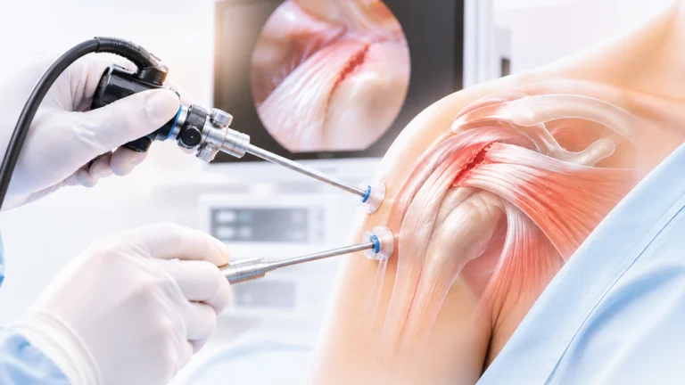
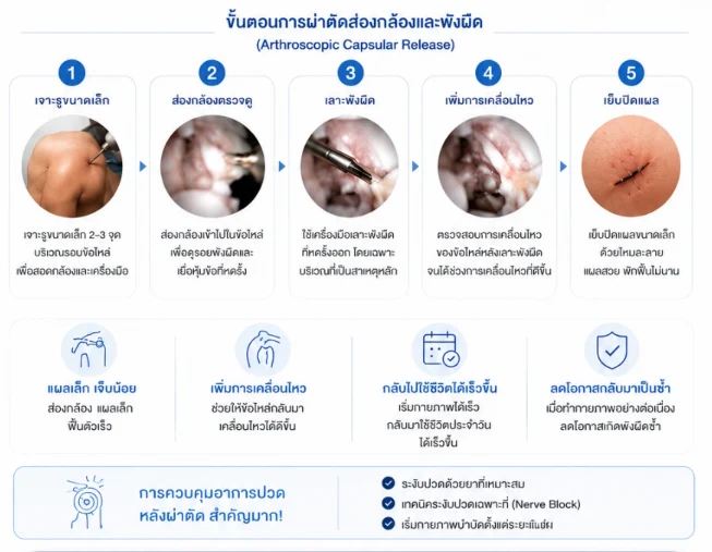
- Operative Procedure: Performed via 2–3 minimally invasive arthroscopic portals. Under direct visualization, hypertrophic fibrotic tissue is released, with focal debridement of the contracted rotator interval and coracohumeral ligament (CHL). Passive ROM is manipulated and confirmed intraoperatively before wound closure with absorbable sutures.
- Clinical Advantages: Minimal portal incisions, low post-surgical morbidity, negligible intraoperative blood loss, precise visualization of concurrent intra-articular pathology, and accelerated recovery timelines.
- Postoperative Protocol: Immediate and uninterrupted postoperative physical therapy is vital to prevent secondary recollapse and capsular re-adhesion. Regional anesthesia (e.g., ultrasound-guided regional nerve blocks) is routinely utilized for post-procedural pain management to facilitate early, pain-free glenohumeral mobilization.
Frequently Asked Questions (FAQ)
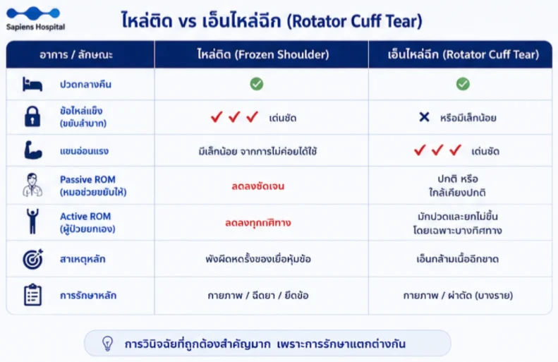
Comparative Clinical Matrix: Frozen Shoulder (Adhesive Capsulitis) vs. Rotator Cuff Tear
| Clinical Parameter / Feature | Frozen Shoulder (Adhesive Capsulitis) | Rotator Cuff Tear |
| Nocturnal Pain | Highly pronounced and persistent | Present (mild to moderate severity) |
| Joint Stiffness / Rigidity | Severe and prominent across planes | Absent or minimal capsular tightness |
| True Motor Weakness | Mild (secondary to disuse atrophy) | Highly pronounced (inability to actively elevate or hold position) |
| Passive ROM (Clinician-Assisted) | Globally restricted in all planes (rigid end-feel even with clinician assistance) | Normal or near-normal (examiner can passively elevate the limb fully) |
| Active ROM (Patient-Initiated) | Globally restricted in all movement planes | Typically restricted only in specific planes corresponding to torn tendons (painful arc) |
| Primary Etiopathology | Synovial inflammation and capsular contracture/fibrosis | Structural tendon tear/avulsion of cuff musculature |
| Primary Treatment Focus | Rehabilitation / Anti-inflammatory Injections / Hydrodilatation / ACR | Rehabilitation / Arthroscopic Rotator Cuff Repair |
- How frequently should physical rehabilitation be performed?: Guided sessions under an orthopedic physical therapist are typically recommended 1–3 times weekly, integrated with daily, diligent home-based range-of-motion stretching exercises.
Conclusion
Frozen shoulder is not simply an isolated, benign mechanical stiffening, but an active inflammatory and fibrotic condition linked with systemic metabolic health. Management should move past passive watchful waiting in favor of proactive, stage-matched clinical evaluations, effective pain modulation, calibrated physical therapy, and timely interventional procedures—such as ultrasound-guided injections, hydrodilatation, or targeted arthroscopic capsular release—to restore functional range of motion and preserve long-term quality of life.
References
- Millar NL, Meakins A, Struyf F, et al. Frozen Shoulder. Nature Reviews Disease Primers. 2022;8:59. doi:10.1038/s41572-022-00386-2.
- Achilova F, Daher M, Nassar JE, et al. Frozen Shoulder: Diagnosis and Treatment of Adhesive Capsulitis. The American Journal of Medicine. 2026;139(5). doi:10.1016/j.amjmed.2026.01.021.
- Navarro-Ledesma S, Luque-Suarez A, et al. A New Perspective of Frozen Shoulder Pathology: The Role of Systemic Inflammation, Fibrosis and Metabolic Dysfunction. Frontiers in Physiology. 2024.
- Drakes S, Aboulhosn P, Pham Y, et al. Adhesive Capsulitis: Review of Current Concepts. Current Physical Medicine and Rehabilitation Reports. 2025. doi:10.1007/s40141-025-00516-3.
- Fernandes MR, et al. Adhesive Capsulitis: Current Concepts. Current Reviews in Musculoskeletal Medicine. 2025.
- Chen MH, Chen CH, Wang KC, et al. A Narrative Review of Adhesive Capsulitis with Diabetes. Journal of Clinical Medicine. 2024;13(19):5696.
- Hernigou P, Deschamps G, Delambre J, et al. Association Between Diabetes Mellitus and Adhesive Capsulitis: A Systematic Review and Meta-analysis. International Orthopaedics. 2026.
- Kim JY, et al. Basic Science Research in Frozen Shoulder: Current Updates. Clinics in Shoulder and Elbow. 2024.
- Karaağaç SS, et al. Quantitative MRI Evaluation of Coracohumeral Ligament and Inferior Glenohumeral Capsule in Adhesive Capsulitis. 2026.
- Le HV, Lee SJ, Nazarian A, Rodriguez EK. Adhesive Capsulitis of the Shoulder: Review of Pathophysiology and Current Clinical Treatments. Shoulder & Elbow. 2017;9(2):75-84.
- Lee BC, et al. Clinical Practice Guidelines for the Diagnosis and Non-Surgical Management of Frozen Shoulder. Annals of Rehabilitation Medicine. 2025.
- Li M, Zhang Y, et al. A Retrospective Comparative Study on the Effectiveness of Multisite Injection and Arthroscopic Capsular Release in Frozen Shoulder. Scientific Reports. 2024.
- Rohit V, et al. A Comparative Study Between Arthroscopic Anterior Capsular Release and Pan-Capsular Release Among Patients With Adhesive Capsulitis. Journal of Arthroscopic Surgery and Sports Medicine. 2026.
- Palacios-Díaz L, et al. Is It Safe to Perform a Full Circumferential Arthroscopic Capsular Release for Frozen Shoulder? A Systematic Review. JSES Reviews, Reports & Techniques. 2026.
- Kim JY, et al. Frozen Shoulder: A Narrative Review of Current Treatment Strategies. Clinics in Shoulder and Elbow. 2025.