 English
 ไทย
 မြန်မာ
 中文
 العربية
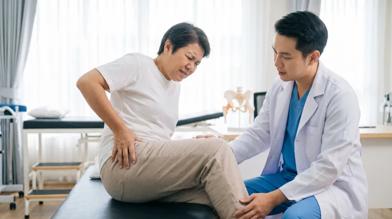
Many individuals suffer from chronic groin or hip pain, particularly when standing up from a chair, climbing stairs, sitting in a car for extended periods, or exercising[cite: 1]. Some are diagnosed with muscle strain, tendon tightness, or hip overuse, yet standard treatments fail to relieve their symptoms[cite: 1].
In certain cases, patients experience distinct mechanical symptoms, such as an audible or palpable “clicking” or “popping” sound during movement, a sensation of intra-articular catching, intermittent locking of the hip, deep groin pain upon hip flexion, or unusual difficulty putting on shoes, socks, and getting into a car[cite: 1].
These symptoms may indicate an Acetabular Labral Tear (Hip Labral Tear), which is a major cause of hip pain among young adults, athletes, and individuals with underlying Femoroacetabular Impingement (FAI)[cite: 1].
Although not as widely recognized by the general public as a knee meniscus tear or a herniated disc, this condition is increasingly identified today due to advancements in MRI diagnostics and hip arthroscopy, revealing that a substantial number of patients with chronic groin pain are indeed suffering from an injured hip labrum[cite: 1].
What Is the Acetabular Labrum?
The hip labrum, or acetabular labrum, is a ring of fibrocartilaginous tissue that surrounds the rim of the acetabular socket[cite: 1]. It functions like a protective “rubber gasket” that deepens the socket, securing the femoral head firmly in place[cite: 1].
The labrum is far more than just a small intra-articular structure; it plays critical biomechanical roles, including enhancing hip joint stability, distributing mechanical loads, preserving the synovial fluid suction seal, and reducing contact stress across the articular cartilage[cite: 1].
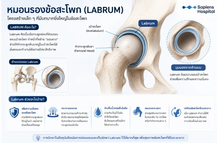
Key Biomechanical Functions of the Labrum
- Enhances Joint Stability: Deepens the socket to stabilize the femoral head, mitigating the risk of micro-instability and subluxation[cite: 1].
- Distributes Load and Contact Pressure: Evenly disperses mechanical forces across the articular surface, preventing localized stress peaks and cartilage degradation[cite: 1].
- Shock Absorption: Dampens impact and shear forces generated during walking, running, jumping, and repetitive dynamic movements[cite: 1].
- Preserves Synovial Fluid (Suction Seal): Maintains an airtight fluid seal that seals synovial fluid within the central compartment, ensuring low-friction articulation and joint lubrication[cite: 1].
- Long-term Joint Protection: A biomechanically sound labrum protects articular cartilage and significantly reduces the long-term risk of developing secondary hip osteoarthritis[cite: 1].
Biomechanical studies have demonstrated that when the labrum loses its integrity or is excised, intra-articular contact stress increases considerably, which can compromise long-term joint longevity[cite: 1]. Consequently, contemporary surgical paradigms have shifted away from simple debridement toward arthroscopic labral repair and preservation whenever feasible[cite: 1].
What Are the Clinical Symptoms of a Labral Tear?
Clinical presentation varies considerably; some individuals experience only mild discomfort, whereas others suffer from severe, debilitating pain that impairs gait, athletic performance, and activities of daily living (ADLs)[cite: 1].
- Deep Groin Pain or Anterior Hip Pain: The most hallmark clinical sign[cite: 1]. Patients typically describe a deep, intra-articular ache rather than superficial muscular soreness[cite: 1]. Pain is characteristically provoked or exacerbated by weight-bearing activities, prolonged sitting, or athletic exertion[cite: 1].
- Referred Pain: Pain may radiate down the anterior thigh, lateral peritrochanteric region, buttock, or medial thigh, occasionally leading to diagnostic confusion with lumbar spine or knee pathology[cite: 1].
- Clicking, Snapping, or Popping: Noticeable particularly upon rising from a seated position, pivoting, getting into a vehicle, or altering posture, often described as an internal mechanical catch or an abrupt loss of smooth joint motion[cite: 1].
- Mechanical Catching or Locking: Sensation of impingement or obstruction during hip flexion, leading to terminal range-of-motion restrictions or episodic joint locking[cite: 1].
- Pain with Prolonged Sitting or Hip Flexion: Aggravated by long car rides, desk work, deep squatting, donning shoes/socks, forward bending, or ascending and descending stairs[cite: 1].
When to Consult an Orthopedic Specialist: Persistent groin pain lasting longer than 6 weeks, painful joint clicking, definitive mechanical catching/locking sensations, or symptoms that fail to resolve with rest and standard conservative care[cite: 1].
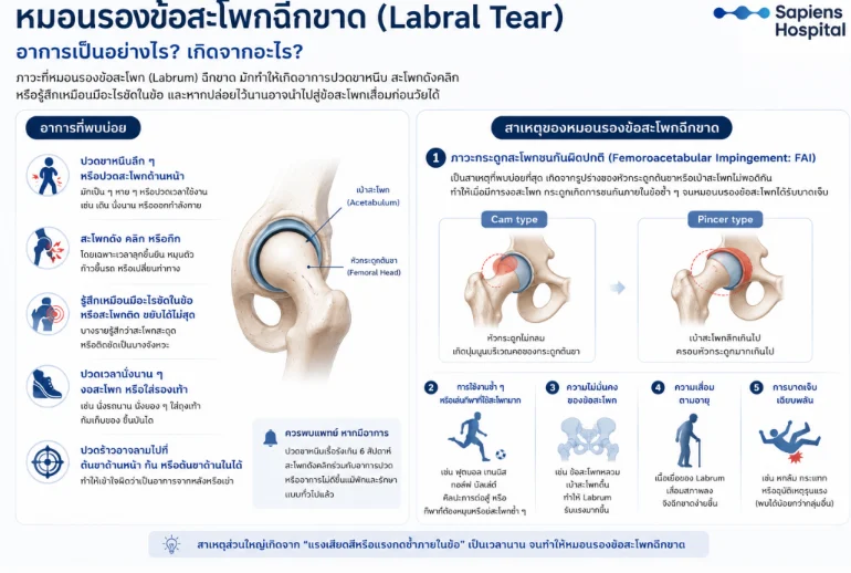
Why Does a Hip Labral Tear Occur? (Etiology)
While commonly assumed to result strictly from high-energy acute trauma, many patients report no preceding history of falls, impacts, or major injuries[cite: 1]. In the majority of cases, the underlying etiology involves “repetitive microtrauma, abnormal shear forces, and chronic intra-articular impingement” over time[cite: 1].
- Femoroacetabular Impingement (FAI): The leading predisposing factor[cite: 1]. Morphological abnormalities of the femoral head-neck junction or acetabular rim cause premature osseous contact during hip motion (particularly flexion and internal rotation), subjecting the labrum to repetitive shear stresses and subsequent tearing[cite: 1].
- Cam Morphology: Asphericity of the femoral head characterized by an osseous prominence (bump) at the anterolateral femoral head-neck junction[cite: 1].
- Pincer Morphology: Over-coverage of the femoral head due to a deep acetabulum (coxa profunda) or focal acetabular retroversion[cite: 1].
- Repetitive Microtrauma and High-Demand Sports: Frequent participation in soccer, tennis, golf, ballet, martial arts, or activities demanding repetitive pivoting, cutting, twisting, and deep end-range hip flexion[cite: 1].
- Hip Joint Instability: Generalized ligamentous laxity or developmental acetabular dysplasia, which places excessive compensatory loading on the labrum[cite: 1].
- Age-Related Tissue Degeneration: Gradual wear, micro-structural breakdown, and loss of tissue elasticity, predisposing the labrum to tear under physiological loads[cite: 1].
- Acute Macrotrauma: High-impact falls, motor vehicle accidents, or sudden violent hip subluxation/dislocation events (comparatively less common than chronic mechanical etiologies)[cite: 1].
How Is a Hip Labral Tear Diagnosed?
An accurate diagnosis requires a comprehensive evaluation correlating clinical history, specialized physical examination maneuvers, and high-resolution imaging modalities[cite: 1]:
- Plain Radiographs (X-ray): Cannot visualize the fibrocartilaginous labral tear directly, but are essential to evaluate bony morphology, assess joint space width, and identify underlying FAI (Cam/Pincer lesions) or acetabular dysplasia[cite: 1].
- Magnetic Resonance Imaging (MRI): Evaluates intra-articular soft tissues, labral tissue pathology, articular cartilage integrity, and periarticular musculotendinous structures[cite: 1].
- MR Arthrography (MRA): Involves intra-articular injection of a dilute gadolinium-based contrast agent prior to scanning, significantly enhancing diagnostic accuracy for subtle, non-displaced labral tears and chondral delamination (performed selectively based on the specialist’s clinical evaluation)[cite: 1].
Does an MRI Diagnosis of a Labral Tear Mandate Surgery? Can It Heal on Its Own?
- Surgery Is Not Universally Required: A substantial number of asymptomatic individuals exhibit labral tears on MRI[cite: 1]. Clinical management adheres strictly to the rule: “Treat the patient, not the imaging study alone”[cite: 1].
- Can It Heal Spontaneously?: Due to the watershed avascular zone of the fibrocartilage, complete biological healing back to pristine histology is rare; however, many patients achieve excellent symptomatic resolution through conservative management[cite: 1]. Pain is predominantly driven by joint inflammation, dynamic overload, and periarticular neuromuscular deficits, all of which are responsive to non-operative interventions[cite: 1].
Conservative (Non-Operative) Treatment Protocol
- Activity Modification: Avoiding deep squats, terminal hip flexion, rapid rotational torque, and aggravating athletic drills[cite: 1].
- Targeted Physical Therapy: Strengthening pelvic and gluteal musculature, restoring core stability, neuromuscular retraining, optimizing movement mechanics, and attenuating focal hip joint contact stresses[cite: 1].
- Pharmacotherapy: Utilization of non-steroidal anti-inflammatory drugs (NSAIDs) to alleviate synovitis and pain, facilitating active participation in physical rehabilitation[cite: 1].
(Patients most likely to succeed with conservative treatment include those without advanced chondral wear, severe joint degeneration, or frequent mechanical locking episodes)[cite: 1].
Indications for Surgical Intervention
Surgical intervention is indicated under the following clinical criteria[cite: 1]:
- Persistent, debilitating pain interfering with ADLs that remains refractory to at least 6 to 12 weeks of structured conservative therapy[cite: 1].
- Prominent mechanical symptoms (recurrent locking, catching, or giving way)[cite: 1].
- Underlying FAI requiring concurrent osseous reshaping (femoroplasty/acetabuloplasty) alongside labral repair[cite: 1].
- High-demand athletes desiring to return to competitive sports[cite: 1].
Arthroscopic Labral Repair (Hip Arthroscopy)
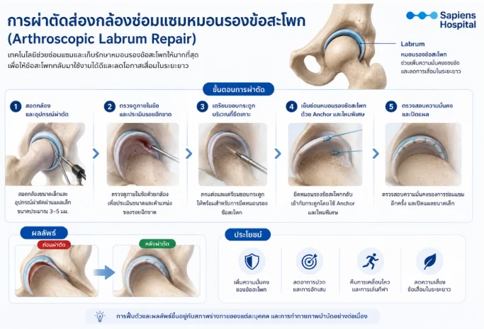
Contemporary surgical management is performed using minimally invasive Hip Arthroscopy through small 3–5 mm portals, centered around the guiding surgical philosophy: “Preserve the Labrum Whenever Possible”[cite: 1].
Surgical Procedure:
- Establishment of arthroscopic portals and introduction of specialized miniature optical instruments and radiofrequency probes[cite: 1].
- Comprehensive diagnostic intra-articular inspection to assess the pattern, extent, and instability of the labral tear, as well as adjacent chondral surfaces[cite: 1].
- Careful rim preparation and decortication of the acetabular bone to establish a bleeding vascular bed conducive to tissue healing[cite: 1].
- Refixation of the detached labrum back to the acetabular rim using bioabsorbable or knotless Suture Anchors with high-tensile sutures[cite: 1].
- Dynamic intraoperative testing under direct visualization to confirm restoration of the labral suction seal and dynamic joint stability, followed by portal closure[cite: 1].
Clinical Benefits: Restores joint stability, alleviates intra-articular pain and synovitis, re-establishes physiological range of motion, facilitates return to sport, and protects articular surfaces against long-term degeneration[cite: 1]. (In instances of extensive tissue irreparable fraying or severe degeneration, debridement or segmental/circumferential labral reconstruction using autograft or allograft may be indicated)[cite: 1].
What Are the Risks of Leaving a Labral Tear Untreated?
If a torn labrum is subjected to ongoing mechanical friction and shear stresses, symptoms typically progress[cite: 1]. Patients face escalating groin pain, worsening sitting intolerance, and loss of athletic capacity[cite: 1]. Moreover, in the presence of unaddressed FAI, untreated labral pathology can accelerate articular cartilage delamination, significantly heightening the risk of early-onset hip osteoarthritis[cite: 1].
Postoperative Rehabilitation and Recovery Timeline
Most patients are mobilized on the day of or the day following surgery, utilizing crutches temporarily to protect the repaired tissue and offload the hip joint[cite: 1]. Sedentary and office-based workers generally resume light duties within 1 to 2 weeks[cite: 1].
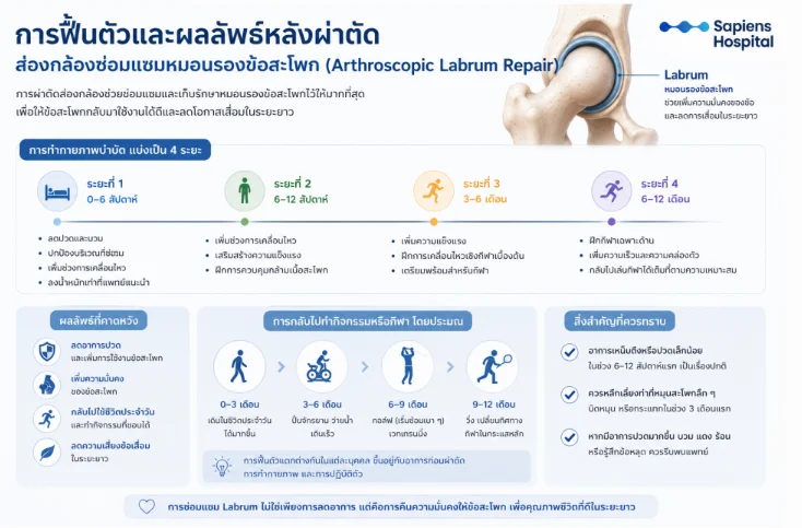
Phased Postoperative Rehabilitation (4 Phases):
- Phase 1 (Weeks 0–6): Pain and edema management, tissue protection, early passive range of motion (PROM) within prescribed safety arcs, and adherence to partial weight-bearing guidelines[cite: 1].
- Phase 2 (Weeks 6–12): Restoration of full active range of motion, progressive gluteal and core strengthening, neuromuscular re-education, and progression toward unassisted reciprocal gait[cite: 1].
- Phase 3 (Months 3–6): Advanced functional resistance training, light plyometrics, sports-specific dynamic patterns, and preparation for heavier physical demands[cite: 1].
- Phase 4 (Months 6–12): High-level agility, speed drills, cutting maneuvers, sport-specific conditioning, and unrestricted return to competitive play upon clinical clearance[cite: 1].
Estimated Timeline for Resuming Physical Activities:
- 0–3 Months: Normalized unassisted walking and progressive activities of daily living[cite: 1].
- 3–6 Months: Stationary cycling, swimming, and brisk treadmill/outdoor walking[cite: 1].
- 6–9 Months: Golf (light rotational swings), progressive barbell resistance training[cite: 1].
- 9–12 Months: Straight-line running, cutting/pivoting sports, and contact/high-impact athletics[cite: 1].
Clinical Notes: Mild tightness or transient periarticular discomfort is completely normal during the initial 6 to 12 weeks[cite: 1]. Aggressive hip rotation, forceful torquing, and high-impact loading must be strictly avoided during the first 3 months[cite: 1]. Clinical outcome literature reports a 80–90% success rate regarding substantial pain reduction and functional return following arthroscopic repair[cite: 1].
Key Takeaways for Patients
- Acetabular labral tears are a major underlying cause of chronic groin and hip pain in young adults, active individuals, and athletes with FAI[cite: 1].
- Characteristic symptoms include deep groin aching, clicking, catching, and mechanical discomfort aggravated by hip flexion and prolonged sitting[cite: 1].
- While MRI/MRA is essential for structural diagnosis, the presence of a tear does not automatically mandate surgery[cite: 1].
- A significant proportion of patients improve satisfactorily with comprehensive non-operative rehabilitation[cite: 1].
- When operative intervention is necessary, modern arthroscopic techniques prioritize anatomic labral repair and preservation to ensure long-term joint function and cartilage protection[cite: 1].
References
- Gómez-Verdejo F, Pérez-Prieto D, Gelber PE, et al. Review of Femoroacetabular Impingement Syndrome. EFORT Open Reviews. 2024;9(12):1152-1165.[cite: 1]
- Fortier LM, Briggs KK, Kivlan BR, Philippon MJ. An Updated Review of Femoroacetabular Impingement Syndrome. Orthopedic Reviews. 2022;14(3):37513.[cite: 1]
- American Academy of Orthopaedic Surgeons (AAOS). Femoroacetabular Impingement (FAI). OrthoInfo.[cite: 1]
- Kelly BT, Williams RJ, Philippon MJ. Arthroscopic Labral Repair in the Hip: Surgical Technique and Review of the Literature. Arthroscopy. 2005;21(12):1496-1504.[cite: 1]
- Mercurio AM, Memon M, Simunovic N, et al. Return to Sport After Primary Hip Arthroscopic Surgery and Labral Treatment: Five-Year Outcomes. Arthroscopy. 2025.[cite: 1]