 English
 ไทย
 မြန်မာ
 中文
 العربية
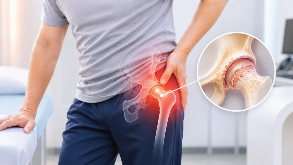
Your doctor diagnosed you with hip osteoarthritis—what should you do next?
Upon receiving a diagnosis of hip osteoarthritis, most patients have similar questions:
- Can hip osteoarthritis be cured?
- Is surgery unavoidable?
- Can I still walk or exercise?
- If I keep walking, will the joint wear down faster?
- How can I prevent my condition from worsening?
The encouraging news is that the majority of patients with hip osteoarthritis do not require immediate surgery. With early, appropriate intervention, many can effectively control pain, maintain ambulatory capacity, continue exercising, and lead fulfilling daily lives for years.
Today, multiple therapeutic pathways are available for managing hip osteoarthritis—ranging from therapeutic exercise, weight control, and physical therapy to pharmacological management, intra-articular injections, and interventional pain procedures, through to total hip replacement for clinically indicated candidates. Therefore, a diagnosis of hip osteoarthritis does not inevitably mean surgery for everyone.
What Is Hip Osteoarthritis?
The hip joint is a major weight-bearing joint that sustains substantial load every time you walk, stand, rise from a chair, negotiate stairs, or even stand still. When articular cartilage progressively wears down, joint space narrows, inflammatory processes develop, and osteophytes (bone spurs) may form—resulting in pain, joint stiffness, and restricted range of motion.
In the early stages, many individuals maintain a functional gait, only noticing discomfort after extended ambulation, when rising from a seated position, or during stair climbing. As disease progression occurs, pain gradually escalates to impact activities of daily living (ADLs)—such as reduced walking tolerance, difficulty putting on shoes or socks, and, in some cases, persistent pain at rest or nocturnal pain disrupting sleep.
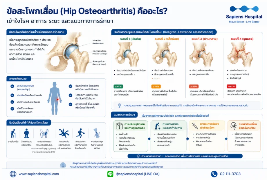
Crucial Clinical Consideration: Radiographic severity does not always correlate directly with clinical symptom severity. Some patients present with pronounced joint degeneration on X-ray yet experience minimal discomfort, whereas others exhibit subtle radiographic alterations alongside debilitating pain. Treatment planning must therefore holistically integrate symptoms, functional lifestyle capacity, and diagnostic imaging findings.
Severity Grading: Kellgren-Lawrence Classification
- Grade 1 (Doubtful/Initial): Minute, questionable joint space narrowing; possible minute osteophytes. Symptoms: Frequently asymptomatic, or mild discomfort following strenuous activity.
- Grade 2 (Mild): Definite osteophytes with possible joint space narrowing. Symptoms: Pain during prolonged walking, ascending/descending stairs, or rising from a chair.
- Grade 3 (Moderate): Multiple moderate osteophytes, definite joint space narrowing, and subchondral sclerosis. Symptoms: Frequent pain, decreased walking distance, and noticeable impairment of routine activities.
- Grade 4 (Severe): Marked joint space narrowing, large osteophytes, severe subchondral sclerosis, and bony contour deformity. Symptoms: Severe pain, significant ambulatory limitation, and frequent resting or nocturnal pain.
Etiology and Risk Factors of Hip Osteoarthritis
- Advanced age
- Overweight or obesity
- Structural hip deformities, including developmental dysplasia of the hip (Hip Dysplasia)
- Femoroacetabular impingement (FAI)
- Previous trauma or intra-articular fractures
- Prolonged participation in high-impact sports or heavy occupational joint loading
- Avascular necrosis (AVN) of the femoral head
Clinical Symptoms of Hip Osteoarthritis
Symptoms typically develop insidiously and worsen progressively, presenting as:
- Groin pain (the most common clinical presentation)
- Gluteal or posterior hip pain
- Referred pain radiating to the anterior thigh or down to the knee
- Decreased walking distance or noticeable antalgic gait (limping)
- Hip joint stiffness, particularly after prolonged sitting or upon morning awakening
- Difficulty donning footwear, putting on socks, or trimming toenails
- Difficulty rising from chairs, climbing stairs, or entering/exiting vehicles
Non-Surgical (Conservative) Management of Hip Osteoarthritis
The therapeutic goal is not solely analgesic symptom control, but restoring functional mobility, maintaining independent daily living, and attenuating radiographic and symptomatic disease progression.
1. Therapeutic Exercise: The Primary Pillar of Conservative Care
Structured exercise strengthens peri-articular musculature, reduces joint contact stress, improves pelvic and gait stability, and alleviates pain levels.
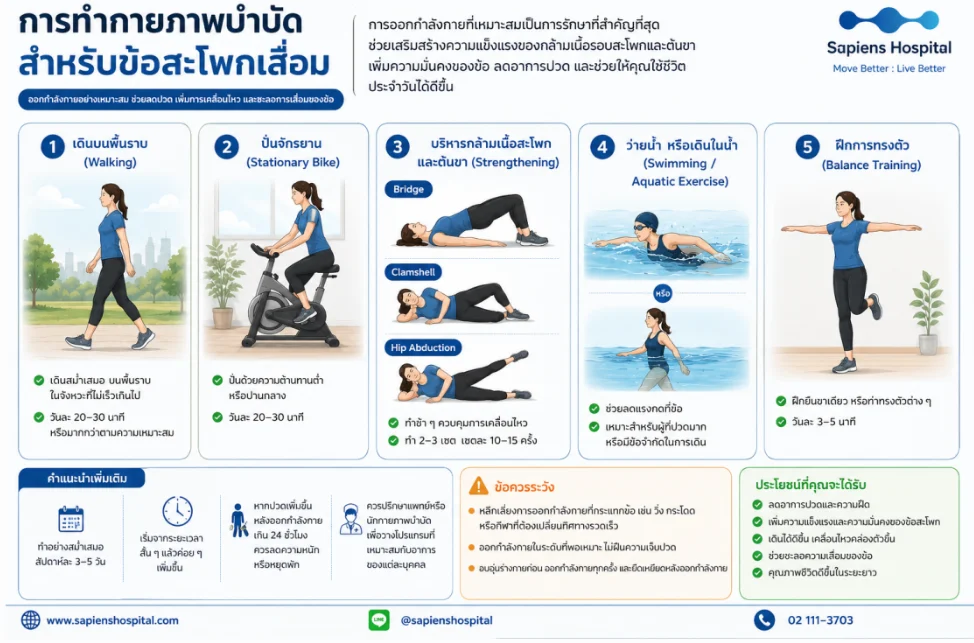
- Recommended Activities: Level-ground walking (20–30 minutes daily), stationary cycling (low to moderate resistance), swimming or hydrotherapy, and balance training (single-leg stance for 3–5 minutes).
- Hip and Thigh Muscle Conditioning: Glute bridge, clamshell, and hip abduction exercises (perform slowly; 2–3 sets of 10–15 repetitions).
- Precautions: Avoid high-impact loading such as running, jumping, or sports requiring abrupt multidirectional cutting. If post-exercise soreness persists beyond 24 hours, decrease exercise intensity.
2. Weight Control and Reduction
During normal ambulation, the hip joint experiences biomechanical loads several times body weight. A reduction of 5% to 10% of total body weight substantially attenuates intra-articular compressive force, diminishes mechanical pain, and delivers clinically meaningful improvements in physical function.
3. Daily Behavioral and Ergonomic Modifications
- Avoid uninterrupted, prolonged standing.
- Pace walking activities with scheduled rest intervals.
- Avoid heavy manual lifting and axial loading.
- Shift posture every 30–60 minutes; avoid sustained static sitting.
- Wear supportive, well-cushioned footwear designed for effective shock absorption.
4. Physical Therapy
A physical therapist conducts targeted assessments of muscle strength, passive and active range of motion, and gait mechanics to prescribe patient-tailored exercise regimens, manual stretch therapies for tight soft-tissue structures, and proper biomechanical instruction for sit-to-stand transitions and stair navigation to support long-term home adherence.
5. Pharmacological Management
- Paracetamol (Acetaminophen): Appropriate for mild persistent pain, maintaining an established safety profile when administered within standard clinical dose limits.
- Non-Steroidal Anti-Inflammatory Drugs (NSAIDs): Such as celecoxib, etoricoxib, naproxen, or ibuprofen; effective in controlling joint pain and synovitis (recommended under medical supervision, with careful monitoring for gastrointestinal, renal, and cardiovascular risks).
6. Assistive Ambulation Devices (Canes)
A cane should be held in the hand contralateral (opposite) to the symptomatic hip to effectively offload joint reaction forces and reduce biomechanical stress across the affected joint.
Intra-Articular Injections and Interventional Procedures
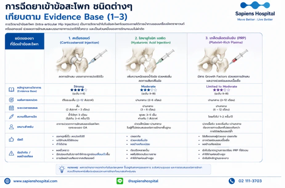
Intra-Articular Hip Injections (Comparative Evidence Base)
| Injectable Agent (Hip Osteoarthritis) | Scientific Evidence Base | Efficacy and Duration | Recommended Frequency | Clinical Advantages & Limitations |
| 1. Corticosteroids | Strong ★★★★★ (Level I–II) | Favorable short-term relief (2 weeks – 3 months) | Repeatable every 3 months (maximum 3–4 times/year) | Rapid analgesic and anti-inflammatory action; however, effects are transient, and repeated frequent administration may accelerate chondral loss. |
| 2. Hyaluronic Acid (HA) | Moderate ★★★☆☆ (Level I–III) | Intermediate relief (3 – 6 months) | Course of 3–5 weekly injections | Provides viscosupplementation; however, clinical efficacy evidence in hip OA remains less conclusive than in knee OA, precluding routine first-line recommendation. |
| 3. Platelet-Rich Plasma (PRP) | Limited to Moderate ★★★☆☆ | Intermediate relief (6 – 12 months) | Typically 1–2 sessions annually | Autologous preparation with favorable safety and bioactive growth factors to modulate joint inflammation; standard preparation protocols remain variable. |
Hip Radiofrequency Ablation (Hip RFA)
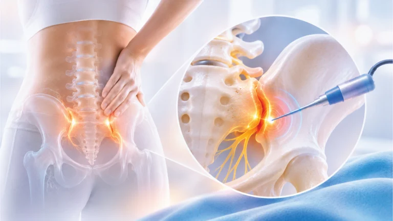
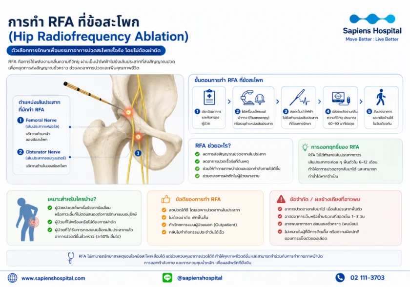
Hip RFA is an interventional modality utilizing radiofrequency thermal energy delivered via specialized needle electrodes to disrupt sensory nerve articular branches transmitting nociceptive pain signals (primarily the articular branches of the femoral nerve anteriorly and the obturator nerve anteromedially).
- Clinical Indications: Chronic hip pain unresponsive to conservative care where surgery is contraindicated, patients presenting with elevated perioperative risk due to medical comorbidities, or those electing to delay definitive arthroplasty.
- Procedure: A diagnostic local anesthetic nerve block is routinely performed first; patients demonstrating $\ge 50\%$ pain reduction proceed to therapeutic radiofrequency ablation.
- Clinical Outcome: Sustained analgesia typically lasting 6 to 12 months (sensory axons gradually regenerate over time, enabling repeat intervention if clinically indicated).
When Should Total Hip Arthroplasty (THA) Be Considered?
Surgical intervention is indicated when pain and joint dysfunction significantly compromise quality of life and conservative pathways have failed to yield satisfactory relief:
- Severe pain severely impeding daily living, ambulation, transfers, or sleep.
- Progressive, marked decline in functional walking tolerance.
- Refractory pain despite structured pharmacological therapy, physical rehabilitation, and interventional injections.
- Moderate to severe radiographic joint destruction (Kellgren-Lawrence grade 3–4) consistent with disabling clinical symptoms.
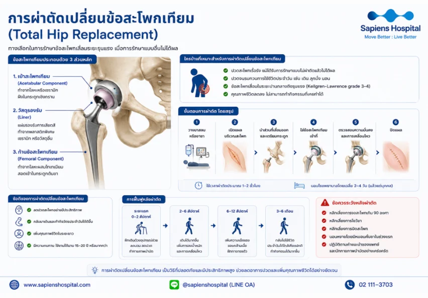
Components of a Total Hip Prosthesis
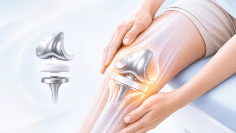
- Acetabular Component (Cup): A metallic or ceramic shell anchored securely within the reamed acetabulum of the pelvis.
- Bearing Liner: An articulating insert fabricated from highly cross-linked polyethylene or ceramic to minimize frictional wear.
- Femoral Component (Stem and Head): A titanium-alloy stem inserted into the femoral canal, coupled with an engineered modular femoral head.
- Clinical Benefits: Profound and predictable pain relief, substantial restoration of independent gait and mobility, and implant longevity extending 15–20 years or longer.
- Postoperative Rehabilitation: Early mobilization initiated within 24 hours post-surgery, mean hospital stay of 2–4 days, and functional return to routine activities within 6–12 weeks.
Summary
Hip osteoarthritis is a prevalent degenerative disorder. While native articular cartilage cannot be fully regenerated, the vast majority of patients can successfully manage symptoms and maintain functional independence for years without immediate surgical intervention.
The core foundation of management encompasses structured exercise, body-weight optimization, targeted peri-articular muscle strengthening, and ergonomic behavioral modifications. If symptoms progress despite conservative measures, image-guided intra-articular injections and hip radiofrequency ablation (Hip RFA) offer effective intermediate interventions prior to considering total hip arthroplasty (THA).
Frequently Asked Questions (FAQ)
- Can hip osteoarthritis resolve on its own?: No, structural cartilage loss does not spontaneously reverse; however, symptoms and pain can be effectively controlled, enabling patients to maintain quality of life for years without surgery through appropriate conservative protocols.
- Can individuals with hip osteoarthritis sit on the floor, cross-legged, or in a kneeling posture?: If these seated postures provoke no discomfort, they are permissible; however, if joint pain or stiffness occurs, prolonged extreme hip flexion and internal rotation should be avoided in favor of ergonomic, standard-height seating.
- What activities should be avoided with hip osteoarthritis?: Avoid heavy lifting, prolonged continuous standing or walking without scheduled rest breaks, and static sitting postures; complete immobilization, however, should be avoided as joint movement is vital for cartilage nutrition.
- Can hip osteoarthritis be managed non-surgically?: Yes, evidence-based non-surgical interventions represent the initial standard of care that should routinely precede surgical consideration in most patients.
References
- American Academy of Orthopaedic Surgeons. Management of Osteoarthritis of the Hip: Evidence-Based Clinical Practice Guideline. Rosemont, IL: American Academy of Orthopaedic Surgeons; 2023.
- Hannon CP, Goodman SM, Austin MS, et al. 2023 American Academy of Orthopaedic Surgeons Management of Osteoarthritis of the Hip Evidence-Based Clinical Practice Guideline. J Am Acad Orthop Surg. 2024;32(10):e544-e556.
- Kolasinski SL, Neogi T, Hochberg MC, et al. 2019 American College of Rheumatology/Arthritis Foundation Guideline for the Management of Osteoarthritis of the Hand, Hip, and Knee. Arthritis Care Res (Hoboken). 2020;72(2):149-162.
- Teirlinck CH, et al. Effect of Exercise Therapy in Patients With Hip Osteoarthritis: A Systematic Review and Cumulative Meta-analysis. Osteoarthritis and Cartilage Open. 2023.
- Mao Y, Qiu B, Wang W, et al. Efficacy of Home-Based Exercise in the Treatment of Pain and Disability at the Hip and Knee in Patients With Osteoarthritis: A Systematic Review and Meta-analysis. BMC Musculoskelet Disord. 2024.
- Belk JW, Kraeutler MJ, Houck DA, et al. Platelet-Rich Plasma Versus Hyaluronic Acid for Hip Osteoarthritis Yields Similarly Beneficial Short-Term Clinical Outcomes: A Systematic Review of Level I and II Randomized Controlled Trials. Arthroscopy. 2022;38(6):2035-2046.
- Pressler MP, et al. Radiofrequency Ablation of the Hip: Review. Ann Palliat Med. 2024.
- Correia R, et al. Ultrasound-Guided Radiofrequency Ablation for Chronic Hip Pain Due to Osteoarthritis. 2024.