 English
 ไทย
 မြန်မာ
 中文
 العربية
Introduction
Hip pain, particularly groin pain, is a prevalent issue affecting both active working professionals and athletes. Symptoms often begin subtly during prolonged sitting, long-distance walking, running, or athletic activities, but gradually intensify over time to interfere with daily life. Some patients experience clicking sounds, mechanical catching when rising from a chair, or sharp pain with hip flexion.
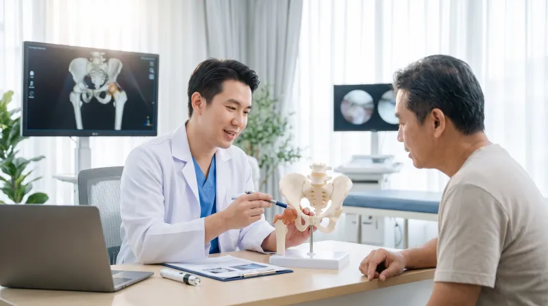
Upon clinical evaluation, a physician may diagnose Femoroacetabular Impingement (FAI) or a Hip Labral Tear, both of which are leading causes of hip joint pain in young and middle-aged adults.
Although initial management typically involves conservative measures—such as activity modification, pharmacotherapy, and targeted physical therapy—hip arthroscopy emerges as an effective surgical option when symptoms persist without satisfactory improvement.
What Is Hip Arthroscopy?
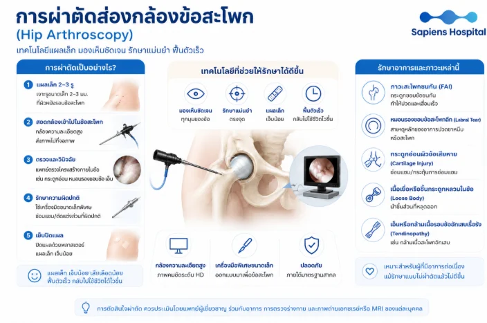
Hip arthroscopy is a minimally invasive surgical procedure utilizing an arthroscope (approximately 4–5 mm in diameter) and specialized surgical instruments inserted through 2–3 small portal incisions (approximately 0.5–1 cm) to inspect, diagnose, and treat intra-articular hip pathologies.
High-definition (HD) endoscopic visualization provides the orthopaedic surgeon with an unobstructed, detailed view of the articular cartilage, acetabular labrum, ligamentum teres, and synovium, allowing concurrent therapeutic intervention to address the root causes of mechanical pain.
- Key Advantages: Minimal incisions, reduced postoperative pain, minimal blood loss, accelerated recovery, and a significantly quicker return to daily routines compared to conventional open hip surgery.
- Treatable Conditions: Femoroacetabular Impingement (FAI), Hip Labral Tears, Chondral/Articular Cartilage Injuries, Intra-articular Loose Bodies, and chronic peri-articular tendinopathy.
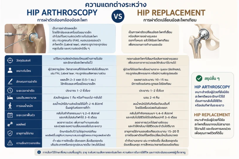
Comparative Overview: Hip Arthroscopy vs. Hip Replacement
| Comparison Parameter | Hip Arthroscopy | Total Hip Replacement (Arthroplasty) |
| Primary Objective | Correct intra-articular anatomical abnormalities and preserve native joint structures (Hip Preservation Surgery) | Replace the severely damaged or degenerated joint with an artificial prosthesis to alleviate pain and restore mobility |
| Ideal Candidates | Young to middle-aged patients without advanced joint degeneration (e.g., FAI, labral tears, focal chondral defects) | Patients with moderate-to-severe osteoarthritis (OA), extensive chondral wear, or significant joint deformities |
| Surgical Approach | Minimally invasive portals (2–3 incisions of 0.5–1 cm) using specialized scopes and micro-instruments | Open incision of approximately 10–15 cm, involving bone resection and prosthetic implant placement |
| Operative Duration | Approximately 1–2 hours | Approximately 1–2 hours |
| Hospital Stay | Typically 1 night or outpatient/day-surgery setting | 2–4 nights of inpatient monitoring |
| Weight Bearing | Partial weight-bearing with crutches for 2–6 weeks (depending on the specific surgical procedure performed) | Immediate or near-immediate weight-bearing with assistive walking devices during early recovery |
| Recovery Timeline | Light activities in 2–6 weeks; return to sports in 3–6 months | Light activities in 4–6 weeks; near-normal daily life in 3–6 months |
| Outcomes & Longevity | Pain reduction, functional restoration, delayed joint degeneration; native joint preservation without implants | Substantial and rapid pain relief; prosthetic implant lifespan of 15–20 years (potential need for revision arthroplasty) |
| Complication Risks | Low (e.g., transient traction-related neuropraxia, infection, localized bleeding) | Low to moderate (e.g., surgical site infection, prosthesis dislocation, deep vein thrombosis, implant loosening) |
Does Arthroscopy Delay Joint Osteoarthritis?: In patients without severe preexisting degeneration, surgical decompression of FAI and labral repair effectively eliminate pathomechanical shear forces, potentially decelerating secondary osteoarthritic progression. However, definitive evidence confirming absolute prevention across all clinical cohorts remains limited.
2. When Should Hip Arthroscopy Be Considered?
Surgical intervention is rarely the primary frontline modality. Patients should initially undertake dedicated conservative therapy, including activity modifications, analgesic and anti-inflammatory medications, pelvic-hip dynamic physical therapy, and diagnostic/therapeutic intra-articular injections. If clinical improvement remains unsatisfactory after 3–6 months of structured non-operative treatment, surgical options should be actively discussed.
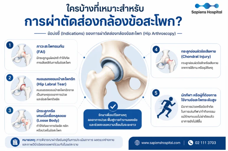
Surgical Indications:
- Femoroacetabular Impingement (FAI): Pathological bony overgrowth at the femoral head-neck junction (Cam) or the acetabular rim (Pincer), causing repetitive abutment and friction during hip flexion, deep squatting, or athletic maneuvers.
- Hip Labral Tear: Persistent joint line pain, mechanical clicking, and catching sensations resulting from torn fibrocartilaginous labral tissue.
- Articular Cartilage / Chondral Injury: Partial-thickness chondral loss or delamination secondary to repetitive impingement or acute mechanical trauma.
- Intra-articular Loose Bodies: Detached osseous or chondral fragments precipitating acute locking, joint entrapment, or audible clunking.
- Athletes and High-Demand Individuals: Patients experiencing recalcitrant pain and mechanical functional limitations impacting athletic performance despite rigorous conservative rehabilitation.
Is an MRI Finding Sufficient on Its Own?: No, it is not sufficient. Asymptomatic morphological variations, including radiological FAI and labral tears, are frequently observed on routine MRI scans in healthy individuals. A definitive surgical decision requires rigorous clinical correlation across three key pillars: Patient Symptoms + Detailed Physical Examination + Diagnostic Imaging (X-rays/MRI), often complemented by an intra-articular local anesthetic diagnostic block to confirm the intra-articular pain generator.
3. Contraindications and Clinical Limitations
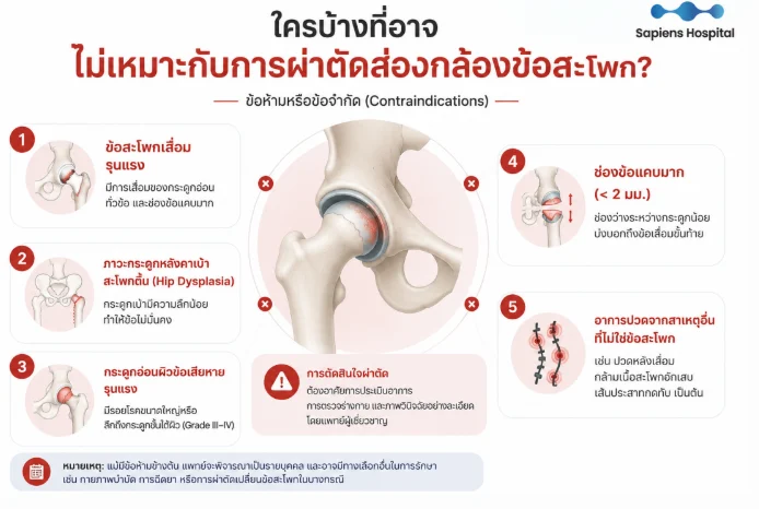
- Severe Hip Osteoarthritis (Advanced OA): Widespread, end-stage chondral collapse cannot be repaired arthroscopically; total hip replacement is the clinically appropriate modality.
- Severely Narrowed Joint Space (< 2 mm): An established radiographic hallmark of advanced joint destruction that correlates with high rates of early conversion to total arthroplasty.
- High-Grade Chondral Lesions (Outerbridge Grade III–IV): Extensive, full-thickness lesions exposing substantial subchondral bone.
- Hip Dysplasia / Structural Joint Instability: Isolated arthroscopic debridement or rim trimming may exacerbate micro-instability and capsule laxity; these conditions require realignment procedures (e.g., Periacetabular Osteotomy – PAO).
- Extraneous Non-Articular Pain Sources: Referred symptoms originating from the lumbar spine, sacroiliac (SI) joint pathology, piriformis syndrome, athletic pubalgia/hernia, or visceral urological/gynecological conditions.
- Age as an Isolated Factor: Advanced age is not an absolute contraindication; the decision relies primarily on the biological quality of articular cartilage and the overall degree of degenerative joint space narrowing.
4. Step-by-Step Surgical Procedure
- Preoperative Setup: Under general anesthesia, the patient is positioned on a specialized traction table. Precise axial traction is applied to distract the central compartment by approximately 1 cm, providing adequate space for safe instrument navigation monitored under continuous fluoroscopy.
- Intraoperative Procedures: Systematic diagnostic arthroscopy is conducted across the central and peripheral compartments, followed by therapeutic interventions as indicated:
- Resection and osteochondroplasty of impinging bone margins (Femoral Osteoplasty / Acetabuloplasty for FAI).
- Refixation and suture anchoring of the labrum (Labral Repair), which demonstrates superior long-term chondroprotective outcomes over simple excision.
- Selective debridement of non-viable, irreparable labral segments.
- Extraction of loose cartilaginous or osseous fragments (Loose Body Removal).
- Operative Time: Routinely ranges between 1–2 hours (complex revision arthroscopies may require extended operating time).
- Postoperative Stay: Most patients require only an overnight stay (1 night) and are cleared for discharge the following day.
5. Postoperative Rehabilitation and Recovery Timeline
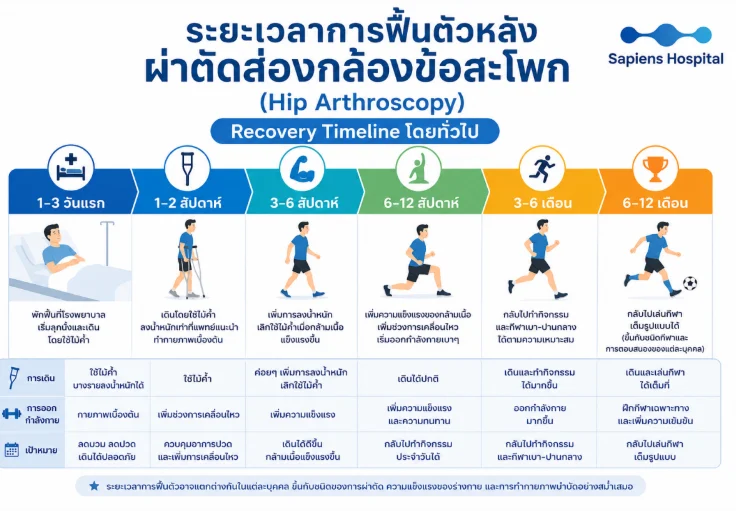
| Recovery Phase | Ambulation & Assistive Devices | Rehabilitation & Exercise Therapy | Primary Clinical Objective |
| Days 1–3 | Early mobilization, sitting up, and protected ambulation with crutches | Gentle bedside isometric and circulation exercises | Edema and pain control; safe transfer mechanics |
| Weeks 1–2 | Crutch-assisted ambulation adhering to weight-bearing restrictions | Passive range of motion (PROM) and gentle pelvic activation | Pain regulation; early hip capsule mobility |
| Weeks 3–6 | Progressive transition toward full weight-bearing; crutch weaning as abductor strength improves | Closed kinetic chain strengthening, low-resistance stationary cycling | Normalization of gait kinematics; core and abductor recruitment |
| Weeks 6–12 | Independent, unassisted physiological ambulation | Progressive neuromuscular resistance training and proprioceptive drills | Full return to unrestricted activities of daily living (ADLs) |
| Months 3–6 | Unrestricted functional walking and stair climbing | Gradual integration into low-impact athletics (e.g., road cycling, swimming) | Cardiorespiratory endurance reconditioning; foundational sports drills |
| Months 6–12 | High-impact athletic mobility and running | Sport-specific multidirectional agility, plyometrics, and full competition drills | Complete functional return to sports; maximized quality of life |
- Return to Work: Sedentary office workers typically resume duties within 2–4 weeks; professions requiring prolonged standing, manual labor, or load carriage require 6–12 weeks.
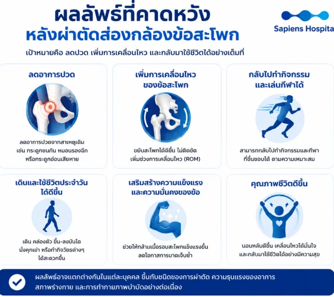
- Clinical Outcomes: The majority of patients achieve significant pain relief, improved range of motion, and successful resumption of athletic performance to pre-injury levels, particularly when treated prior to widespread chondral degeneration.
6. Complications and When to Seek Medical Attention
- Potential Surgical Complications:
- Transient perineal, thigh, or foot paresthesia secondary to mechanical traction neuropraxia, which routinely resolves spontaneously over several days to weeks.
- Localized hematoma, active bleeding, or deep surgical site infection (exceedingly rare).
- Deep Vein Thrombosis (DVT).
- Iatrogenic neurovascular injury or chondral scuffing (extremely rare in experienced hands).
- Recurrence Rate: Symptom recurrence is low when adequate bony resection and structural labral stabilization are achieved, though recurrent issues may stem from new traumatic insult or underlying age-related degeneration.
When to Consult an Orthopaedic Specialist:
- Groin or lateral hip pain persisting continuously for more than 6–12 weeks despite adequate rest and pharmacotherapy.
- Pain exacerbated by prolonged sitting, rising from seated postures, entering/exiting vehicles, walking, running, or athletic pursuits.
- Mechanical clicking, catching, popping, or painful intra-articular locking sensations.
- Progressive limitation in hip internal rotation or flexion range of motion.
- Lack of symptomatic resolution following 3–6 months of compliant conservative physical therapy.
Conclusion
Hip arthroscopy represents a clinically proven, tissue-sparing intervention for patients with symptomatic FAI and labral tears prior to the onset of severe osteoarthritis. The cornerstones of high surgical success include rigorous patient selection, meticulous arthroscopic technique, precise bony contouring, anatomical labral refixation, and adherence to structured, criterion-based rehabilitation protocols.
References
- Suarez-Ahedo C, Camacho-Galindo J, López-Reyes A, et al. A comprehensive review of hip arthroscopy techniques and outcomes. SAGE Open Med. 2024;12:20503121231222212. doi:10.1177/20503121231222212.
- Follet L, Khanduja V, Thevendran G, et al. How to start with hip arthroscopy in a safe and effective manner, using an evidence-based approach. SICOT J. 2024;10:35.
- de SA D, Lian J, Sheean AJ, et al. A Systematic Summary of Systematic Reviews on the Topic of Hip Arthroscopic Surgery. Orthop J Sports Med. 2018;6(9):2325967118796222.
- Pietrzak JRT, Donaldson MJ, Kayani B, et al. Hip Arthroscopy: A Narrative Review of the Current Literature. J Musculoskelet Disord Treat. 2018;4:057.
- Griffin DR, Dickenson EJ, O’Donnell J, et al. Hip Arthroscopy versus Best Conservative Care for the Treatment of Femoroacetabular Impingement Syndrome (UK FASHION): A Multicentre Randomised Controlled Trial. Lancet. 2018;391:2225-2235.
- Palmer AJR, et al. Mid-term Outcomes of Hip Arthroscopy versus Personalised Hip Therapy for Femoroacetabular Impingement Syndrome. Bone Joint J. 2023.
- Monselli C, et al. State of the Art in Rehabilitation Strategies After Hip Arthroscopy for Femoroacetabular Impingement. J Clin Med. 2024.
- Akhtar M, Wen J, Razick D, et al. Outcomes of Arthroscopic Joint Preservation Techniques for Chondral Lesions of the Hip: An Updated Systematic Review. Arthroscopy. 2023.
- Benefits of Hip Arthroscopy Surgery for Soccer Players: A Systematic Review and Meta-analysis. BMC Musculoskelet Disord. 2026;27:38.