 English
 ไทย
 မြန်မာ
 中文
 العربية
To understand the matter.The MRI scan revealed no cause for the pain. From causes and symptoms to diagnosis and treatment approaches, by a team of pain specialists at Sapiens Hospital.
MRI reveals no cause for pain: Overview and medical care guidelines.
I've had an MRI…why haven't I gotten an answer yet?
Limitations of Magnetic Resonance Imaging (MRI) in Chronic Pain Diagnostics
Many patients with chronic pain come into the examination room with MRI films in hand, but are left with crucial questions such as, “Doctor, I've had an MRI, but I still don't know what's wrong?” or “Why have I shown the films to several doctors, but they all give different explanations?” This confusion occurs more frequently than you might think, and the answer doesn't lie with either party's fault, but rather with the “nature and limitations of the MRI machine itself,” which many people often misunderstand.
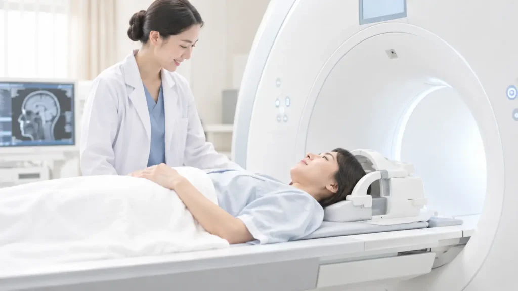
Most patients expect MRI scans to be like a quick answer key, instantly revealing the cause of their pain, whether surgery is needed, and the appropriate treatment. However, in clinical practice, MRI is not designed to directly answer questions about pain; it is merely a tool for capturing anatomical structures within the body. These anatomical images often cannot tell the whole story of pain.
Clinical turning point: Age-related decline detected in the general population.
In the early days of MRI technology's widespread use in the medical field, most doctors believed that high-resolution scans could explain almost all causes of back and neck pain. However, when proactive research was conducted using MRI scans on a “normal population that had never experienced neck or back pain throughout their lives,” the results surprised the medical community greatly.
Several large-scale research studies consistently confirm that seemingly normal individuals with no symptoms can show significant degenerative pathology on MRI scans, such as:
- Degenerative disc disease, intervertebral disc bulging, or disc herniation.
- Spinal hypertrophy or osteoarthritis.
Statistics indicate that these signs of degeneration are commonly found in all humans, especially as they age. Similar to the appearance of wrinkles on the skin or graying hair during the natural aging process (Aging spine), this has led to a major turning point in the field of pain management, making patients realize that “what appears degeneration on MRI scans may not be what is currently causing them pain,” and that these degenerative findings don't always indicate a disease requiring treatment.
The physiological and biomechanical limitations (The Static View Limitation)
The primary reason why MRI images cannot directly reflect a patient's pain level is due to three critical limitations:
1. Scans show only anatomy, but not the inflammatory process (Anatomy vs function).
MRI provides excellent anatomical still images, but it cannot pinpoint which structures on the film are acting as the true “pain generator.” This tool cannot measure pain levels resulting from bodily functions, nor can it pinpoint where minor inflammatory processes are occurring or which peripheral nerves are being irritated/short-circuited during real-world use.
2. Limitations of the lie-down examination position in the tunnel (The Lie-Down Artifact)
MRI scans require patients to lie still on their backs for several minutes to prevent blurred images. However, in daily life, pain often flares up in other postures, such as when standing for long periods, walking long distances, sitting at an office desk continuously, bending over to lift heavy objects, or climbing stairs. Therefore, an MRI image in a supine position is just a single static shot that cannot accurately reflect what is actually happening to the spine as the patient moves in real life.
3. The pressure within the intervertebral disc varies with posture.
Biomechanics research has clearly demonstrated that pressure within the intervertebral discs is lowest in the supine position, and this pressure rapidly increases when changing posture to standing, sitting, or bending forward. This leads to a common clinical phenomenon: some patients experience severe nerve root compression when standing or walking, but when examined on an MRI scan, the reduced pressure causes the disc to collapse back into its normal position, resulting in an MRI image that appears less severe or normal, contrary to the actual pain.
Deep lesions that are often overlooked on MRI scans (Hidden Pain Generators)
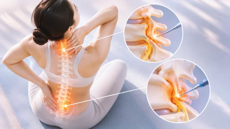
In patients with chronic axial neck pain or chronic axial low back pain without radiating pain to the limbs, MRI scans often show significant limitations and are prone to misdiagnosis. This is because the true origin of the pain is often hidden in deep structures that are not clearly visible on MRI, including:
- Pain from chronic inflammation of the posterior spinal joints (Cervical / Lumbar Facet Joint Pain)
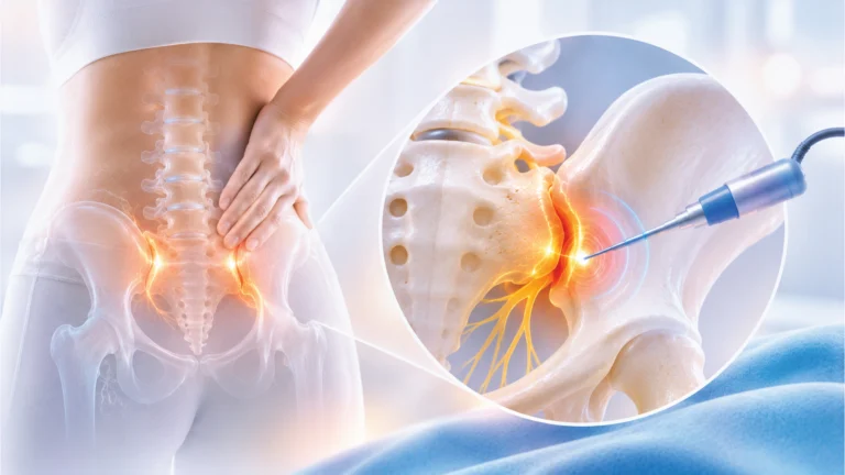
- Pain caused by thickened ligaments or fascia pulling on the posterior pelvic joint (Sacroiliac Joint Pain).
- A minor inflammatory process of the joints, or a condition of deep muscle tissue spasm.
Therefore, each physician may interpret and prioritize the details in an MRI film differently depending on the symptoms associated with the patient at that time.
Our core principle in treatment: “We treat patients, not MRI images.”
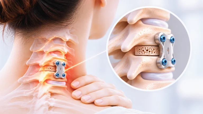
In pain management practice, it's common to encounter cases of patients who have undergone spinal fusion surgery. Subsequent X-ray or CT scans often show technically perfect results, with the bone firmly fused and screws in optimal anatomical positions. However, the patient still asks the same disheartened question: “Why am I still suffering from the same pain?”
This reflects the professional truth that patients come to see a doctor not because of abnormal MRI images, but because of pain that is affecting their quality of life. Therefore, accurate diagnosis and targeted treatment planning must not be based solely on MRI scans, but must utilize a holistic clinical jigsaw puzzle approach, systematically integrating information from various sources, including:
Accurately diagnosing the origin of pain.
Diagnosing the cause of pain, particularly chronic neck and back pain, requires an evaluation of multiple clinical findings and should not rely solely on MRI or radiographic images.
Key elements in diagnosis.
- Accurate diagnosis
- Medical history
- Characteristics and patterns of pain.
- Neurological examination
- X-ray
- Electrical Diagnostics (EMG/NCS)
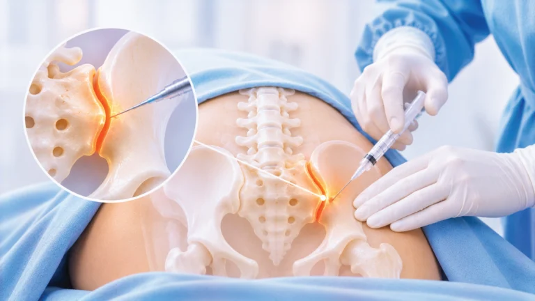
- Diagnostic Block
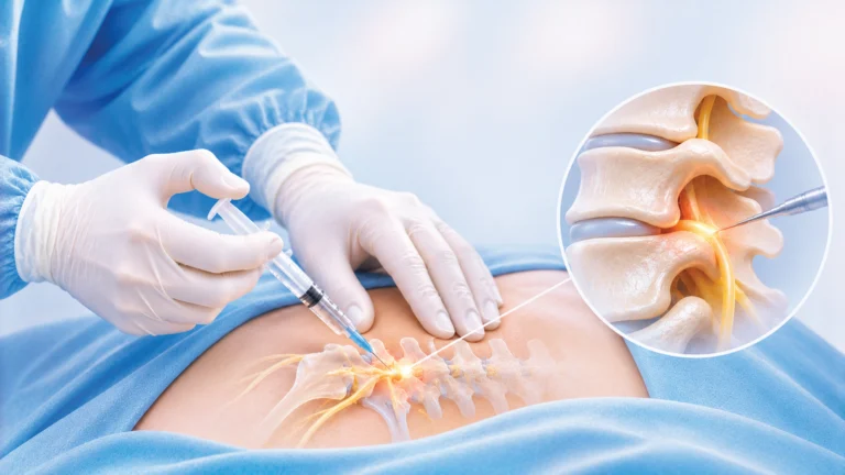
In particular, a diagnostic block (specific local anesthetic injection) is a crucial piece of the puzzle in “temporarily switching off” the pain to identify and confirm the true source of the pain before initiating minimally invasive, targeted, and highly effective long-term treatment.
References - Boden SD, Davis DO, Dina TS, et al. Abnormal Magnetic-Resonance Scans of the Lumbar Spine in Asymptomatic Subjects. J Bone Joint Surg Am. 1990;72:403-408.
- Brinjikji W, Luetmer PH, Comstock B, et al. Systematic Literature Review of Imaging Features of Spinal Degeneration in Asymptomatic Populations. AJNR Am J Neuroradiol. 2015;36:811-816.
- Brinjikji W, Diehn FE, Jarvik JG, et al. MRI Findings of Disc Degeneration are More Prevalent in Adults with Low Back Pain than in Asymptomatic Controls. AJNR Am J Neuroradiol. 2015;36:2394-2399.[cite: 15, 31]
- Wocial K, Feldman BA, Mruk B, et al. Imaging Features of the Aging Spine. Pol J Radiol. 2021;86:e380-e386.
- Wáng YXJ, Wu AM, Ruiz Santiago F, et al. Informed Appropriate Imaging for Low Back Pain Management: A Narrative Review. J Orthop Translat. 2018;15:21-34.
- Del Grande F, Maus TP, Carrino JA. Imaging the Intervertebral Disk: Age-Related Changes, Herniations, and Radicular Pain. Radiol Clin North Am. 2012;50:629-649.
- Pfirrmann CW, Metzdorf A, Zanetti M, et al. Magnetic Resonance Classification of Lumbar Intervertebral Disc Degeneration. Spine. 2001;26:1873-1878.
Diagnostic tests and treatment guidelines, including an MRI scan, did not find the cause of the pain.
A pain specialist will take a medical history, perform a physical examination, and plan a treatment plan appropriate for each patient.
Warning signs that warrant a doctor's visit.
If the symptoms interfere with your daily life or are unusual, you should seek evaluation from a specialist.
Frequently Asked Questions about MRI scans that don't find the cause of pain (FAQ)
How should I prepare before seeing a doctor because an MRI doesn't find the cause of my pain?
It is advisable to record the symptoms, duration of the illness, and previous treatment history to aid in diagnosis.
How long does treatment and rehabilitation take if an MRI doesn't find the cause of the pain?
The recovery time depends on the cause and severity of the condition, and the doctor will continuously monitor the patient's progress.