 English
 ไทย
 မြန်မာ
 中文
 العربية
Chronic hip pain: Do not assume it is just muscle inflammation
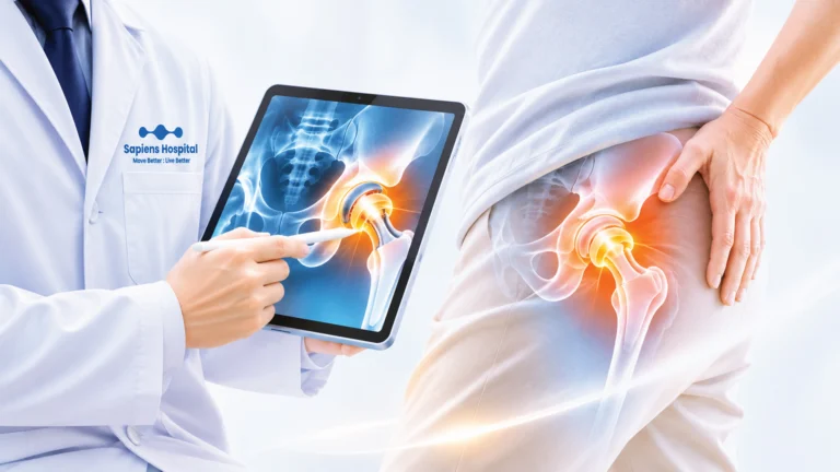
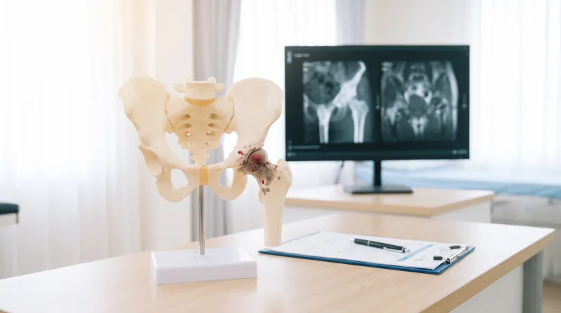
Hip pain is a common clinical complaint[cite: 1]. Many individuals first notice deep groin pain, discomfort during walking, ascending or descending stairs, rising from a chair, or bending to put on shoes[cite: 1]. Consequently, many assume it is merely muscle strain or tendonitis, opting for rest or self-medicating with over-the-counter analgesics[cite: 1].
Although most hip pain does not stem from life-threatening conditions, symptoms persisting for several weeks, reduced walking tolerance, or difficulty with weight-bearing can signal underlying hip joint pathology that requires medical evaluation[cite: 1]. A notable condition is Avascular Necrosis (AVN) of the Hip, also known as Osteonecrosis of the Femoral Head[cite: 1]. In this condition, diminished blood supply to the femoral head causes progressive bone cell death, structural compromise, and potential collapse, ultimately leading to severe secondary osteoarthritis if left untreated[cite: 1].
The encouraging news is that when detected at an early stage—prior to femoral head collapse—effective joint-preserving interventions remain viable for many patients[cite: 1].
What is Avascular Necrosis (AVN) of the Hip?
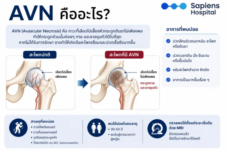
Hip AVN is a condition characterized by inadequate vascular perfusion to the femoral head, leading to progressive death of osteocytes and bone marrow cells[cite: 1]. Deprived of biological repair capacity, the femoral head loses structural integrity, develops subchondral fractures, and eventually collapses, causing articular surface distortion and subsequent hip osteoarthritis[cite: 1].
Disease severity is determined not only by symptomatic pain, but primarily by pathological staging and the degree of femoral head collapse[cite: 1]. Therefore, early diagnosis is essential, as it provides a critical window for joint-preserving strategies aimed at halting disease progression and maintaining the native hip joint[cite: 1].
- Common Symptoms: Deep groin, hip, or anterior thigh pain; exacerbated by walking, prolonged sitting or standing, or climbing stairs; joint stiffness and limited range of motion; progressive worsening over time[cite: 1].
- Common Etiologies: Corticosteroid use, heavy alcohol consumption, trauma/fracture, systemic conditions such as systemic lupus erythematosus (SLE) or sickle cell disease[cite: 1].
- Predominant Age Group: 30–50 years of age (higher prevalence in males than females)[cite: 1].
- Diagnostic Workup: Detectable in early stages via MRI (earlier detection correlates with superior clinical outcomes and joint preservation)[cite: 1].
What Causes Avascular Necrosis of the Hip?
The primary pathogenic mechanism involves disrupted or arrested microvascular blood flow to the femoral head[cite: 1]. This ischemic state deprives osseous tissue of oxygen and essential nutrients, resulting in osteocyte necrosis[cite: 1]. Over time, bone architecture weakens, predisposing it to mechanical collapse[cite: 1].
Etiological factors are categorized into two primary groups[cite: 1]:
- Traumatic AVN:
Arises from acute mechanical trauma that disrupts the arterial blood supply to the femoral head, such as femoral neck fractures, hip dislocations, or severe high-energy impact injuries[cite: 1]. Even after bony union is achieved, microvascular perfusion may fail to re-establish completely, leading to secondary AVN[cite: 1]. - Non-traumatic AVN:
More prevalent, typically associated with systemic medical conditions or risk factors that impair microvascular bone perfusion, including[cite: 1]:
- Prolonged or high-dose systemic corticosteroid therapy[cite: 1]
- Chronic excessive alcohol consumption[cite: 1]
- Systemic Lupus Erythematosus (SLE)[cite: 1]
- Sickle cell disease[cite: 1]
- Coagulation disorders / Hypercoagulability[cite: 1]
- Post-solid organ transplantation[cite: 1]
- Caisson disease (decompression sickness)[cite: 1]
(Approximately 20–30% of cases present with no identifiable cause and are classified as idiopathic AVN)[cite: 1].
Symptoms of Hip Avascular Necrosis
In the earliest stages, patients may remain asymptomatic[cite: 1]. As ischemia progresses, patients experience a deep, aching pain localized to the groin, hip, or buttock, exacerbated by weight-bearing, ambulation, climbing stairs, rising from seated positions, or prolonged standing[cite: 1]. Pain often temporarily improves with rest, frequently leading to misattribution as routine muscular strain[cite: 1].
With clinical progression, pain episodes become more severe and frequent[cite: 1]. Walking tolerance diminishes, an antalgic gait (limping) develops, and active/passive range of motion decreases—particularly internal rotation of the hip[cite: 1].
Upon subchondral collapse, pain occurs with almost every weight-bearing step[cite: 1]. Some patients suffer from severe pain at rest or nocturnal pain that disrupts sleep, substantially impairing quality of life and activities of daily living[cite: 1].
Prompt medical consultation is recommended if experiencing the following:
- Persistent groin or hip pain lasting more than 2–4 weeks[cite: 1]
- Pain upon ambulation or the onset of an antalgic limp[cite: 1]
- Progressive restriction in hip range of motion[cite: 1]
- Intensifying pain during rest or at night[cite: 1]
How is Hip AVN Diagnosed?
Diagnostic evaluation begins with a thorough medical history and physical examination, followed by targeted diagnostic imaging[cite: 1]:
- Plain Radiography (X-ray): Standard baseline imaging to evaluate overall femoral head morphology and joint space[cite: 1]. However, in early pre-collapse stages, plain radiographs are frequently unremarkable despite prominent clinical symptoms[cite: 1].
- Magnetic Resonance Imaging (MRI): The definitive diagnostic gold standard when AVN is clinically suspected but radiographs are normal[cite: 1]. MRI reliably detects ischemic marrow changes prior to structural collapse and precisely evaluates lesion size, location, and geometry to guide management[cite: 1].
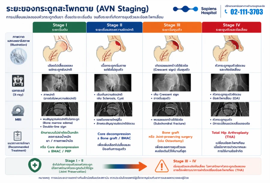
Avascular Necrosis Staging (AVN Staging)
AVN is classified into four stages based on progressive pathological and radiographical changes in the femoral head[cite: 1]:
| Disease Stage | Pathological Characteristics | Radiographic Findings (X-ray) | MRI Findings | Recommended Treatment Approaches |
| Stage I (Early Stage) | Compromised microvascular perfusion; bone architecture remains intact without collapse[cite: 1] | Normal (no visible radiographic abnormalities)[cite: 1] | Bone marrow edema pattern, “double-line sign”[cite: 1] | Primarily non-surgical management, protected weight-bearing, pharmacotherapy/physical therapy, or Core Decompression combined with BMAC/PRP[cite: 1] |
| Stage II (Pre-collapse Structural Alterations) | Osteonecrosis with reactive remodeling/sclerosis; structural spherical contours maintained without collapse[cite: 1] | Demarcated abnormalities: sclerosis, subchondral cysts, or osteopenia[cite: 1] | Enlarging necrotic segment with clear demarcation borders[cite: 1] | Core Decompression combined with Bone Grafting / BMAC to decompress, restore blood supply, and prevent collapse[cite: 1] |
| Stage III (Early Collapse Stage) | Subchondral fracture initiation; early flattening/collapse of the femoral head[cite: 1] | Pathognomonic “Crescent sign”, visible subchondral collapse[cite: 1] | Subchondral fracture line confirmation and focal femoral head flattening[cite: 1] | Bone grafting or joint-preserving osteotomy (e.g., rotational/angular) to redistribute mechanical load and delay degeneration[cite: 1] |
| Stage IV (Advanced Collapse & Osteoarthritis) | Extensive structural femoral head collapse, articular destruction, and joint space obliteration[cite: 1] | Pronounced femoral head collapse and secondary hip osteoarthritis (OA) changes[cite: 1] | Severe head collapse, joint space narrowing, and widespread degenerative changes[cite: 1] | Total Hip Arthroplasty (THA)[cite: 1] |
Clinical Staging & Management Summary:
- Stage I – II: Pre-collapse phase; high probability of successful native joint preservation (Joint Preservation)[cite: 1].
- Stage III – IV: Post-collapse phase with developing or end-stage osteoarthritis; joint preservation efficacy is limited, and Total Hip Arthroplasty (THA) becomes the definitive treatment of choice[cite: 1].
Treatment Strategies by Stage
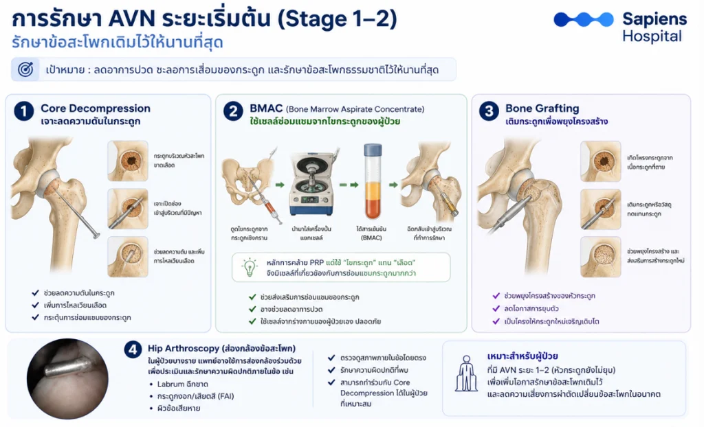
1. Pre-Collapse Treatment (Stage I–II: Joint Preservation)
The primary clinical goal is preserving the native biological joint, retarding disease progression, and delaying or eliminating the future necessity for arthroplasty[cite: 1].
- Core Decompression: A minimally invasive procedure that drills small decompression channels into the necrotic zone to reduce intraosseous pressure, facilitate revascularization, and promote osteogenesis[cite: 1].
- Bone Marrow Aspirate Concentrate (BMAC): Autologous bone marrow harvested from the iliac crest, concentrated via centrifugation to isolate osteoprogenitor and mesenchymal cells, and injected into the necrotic lesion to stimulate osteogenesis[cite: 1].
- Bone Grafting: Introducing autologous, allogeneic, or synthetic bone graft materials into the necrotic cavity to provide structural mechanical support and prevent subchondral collapse[cite: 1].
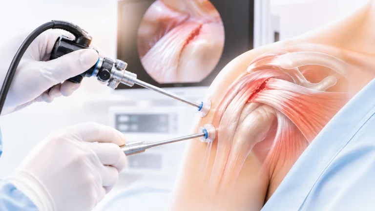
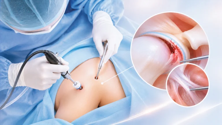
- Hip Arthroscopy: Utilized in select patients for direct intra-articular inspection and management of concomitant pathology, such as acetabular labral tears or femoroacetabular impingement (FAI)[cite: 1].
2. Management of Stage III AVN
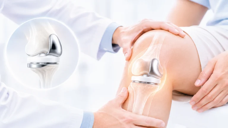
Once subchondral collapse commences, clinical management becomes more complex[cite: 1]. In younger patients with minimal focal collapse, structural bone grafting or realignment osteotomy may be considered to shift weight-bearing load away from the collapsed segment[cite: 1]. However, in cases with substantial collapse, severe intractable pain, or marked functional impairment, total joint arthroplasty delivers superior, more reliable clinical outcomes[cite: 1].
3. Management of Stage IV AVN
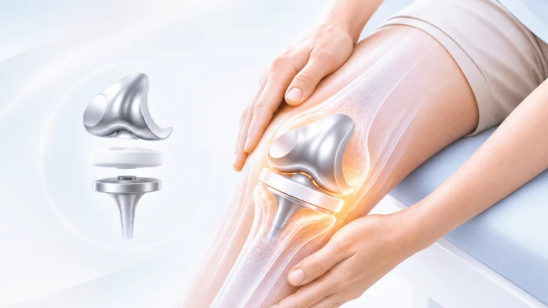
Following extensive collapse and secondary degenerative joint disease, joint-preserving options are no longer viable[cite: 1]. Total Hip Arthroplasty (THA) represents the gold standard intervention in this stage, offering definitive pain relief, restoring functional range of motion, and returning patients to high-quality daily function[cite: 1].
Lifestyle Modifications & Self-Care Guidelines (Living with AVN)
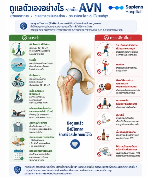
Recommended Practices:
- Pain-Free Walking: Moderate walking for 30–45 minutes daily within a pain-free threshold improves joint lubrication and eases stiffness[cite: 1].
- Swimming: Non-weight-bearing cardiovascular exercise that builds peritrochanteric muscle strength without impact stress on the joint[cite: 1].
- Stationary Cycling: Cycling on level terrain or with low mechanical resistance for 30–45 minutes[cite: 1].
- Elliptical Trainer: Effective low-impact aerobic exercise[cite: 1].
- Hip Abductor and Core Strengthening: Supports pelvic stability, unloads excessive joint pressure, and improves gait mechanics[cite: 1].
- Body Weight Optimization: Every kilogram of weight reduction cumulatively alleviates mechanical compressive loads across the hip during ambulation[cite: 1].
- Balanced Bone Nutrition: Adequate dietary protein, vitamin D, and calcium intake to support ongoing bone metabolism[cite: 1].
- Regular Clinical Follow-ups: Periodic evaluations to monitor radiographic progression and adapt therapeutic plans promptly[cite: 1].
Activities to Avoid:
- Running, jogging, or high-impact aerobics[cite: 1]
- Jumping and high-velocity plyometric exercises[cite: 1]
- Contact and high-torque sports (e.g., soccer, basketball, tennis)[cite: 1]
- Heavy lifting or excessive axial loading[cite: 1]
- Tobacco smoking (causes peripheral vasoconstriction, reduces bone perfusion, and impairs osteogenic repair)[cite: 1]
- Heavy alcohol consumption[cite: 1]
- Unsupervised, prolonged, or illicit self-administration of corticosteroids[cite: 1]
Conclusion
Avascular Necrosis (AVN) of the hip is a condition caused by compromised blood supply to the femoral head, leading to structural osteonecrosis, potential collapse, and secondary hip osteoarthritis[cite: 1]. Early detection represents the single most important determinant of success, providing a window to deploy joint-preserving therapies and protect the native joint[cite: 1]. In advanced stages where structural collapse has occurred, Total Hip Arthroplasty (THA) remains an exceptionally effective, evidence-based surgical procedure that eliminates chronic pain and reliably restores functional mobility[cite: 1].
Frequently Asked Questions (FAQ)
- Can I engage in walking exercise if diagnosed with hip AVN?: Yes, provided you are in the early stages and walking does not induce or intensify pain[cite: 1]. Emphasize low-impact activities like swimming, stationary cycling, or controlled flat-ground walking, and avoid high-impact athletic pursuits[cite: 1].
- What is the typical age of onset for hip AVN?: While it can present across various age groups, it predominantly affects young and middle-aged adults between 30 and 50 years of age[cite: 1].
- Can AVN affect both hip joints simultaneously?: Yes[cite: 1]. Bilateral involvement is common, especially in individuals with systemic corticosteroid exposure, heavy alcohol intake, or SLE[cite: 1]. A patient may present with symptoms on only one side while MRI demonstrates early-stage asymptomatic involvement in the contralateral hip[cite: 1].
- Can nutritional supplements or vitamins cure hip AVN?: To date, there is no clinical evidence demonstrating that collagen, glucosamine, or vitamin supplements can arrest or reverse the pathology of AVN[cite: 1]. Nevertheless, clinicians frequently recommend supplemental vitamin D and calcium to optimize generalized bone metabolic health[cite: 1].
References
- Mont MA, Salem HS, Piuzzi NS, Goodman SB, Jones LC. Nontraumatic osteonecrosis of the femoral head: Where do we stand today? J Bone Joint Surg Am. 2020;102(12):1084-1099.[cite: 1]
- Lieberman JR, Berry DJ, Mont MA, Aaron RK, Callaghan JJ, Rajadhyaksha AD, et al. Osteonecrosis of the hip: Management in the twenty-first century. Instr Course Lect. 2003;52:337-355.[cite: 1]
- Yoon BH, Jones LC, Chen CH, Cheng EY, Cui Q, Drescher W, et al. Etiologic classification criteria of ARCO on femoral head osteonecrosis. J Arthroplasty. 2019;34(1):163-168.[cite: 1]
- Association Research Circulation Osseous (ARCO). ARCO Consensus on the staging and management of osteonecrosis of the femoral head. J Orthop Res. 2019.[cite: 1]
- Pierrepont JW, Marel E, Baré JV, et al. Management of osteonecrosis of the femoral head: Current concepts. Bone Joint J. 2024;106-B(Suppl):3-12.[cite: 1]
- American Academy of Orthopaedic Surgeons (AAOS). Osteonecrosis of the Hip (Avascular Necrosis). Updated 2024.[cite: 1]
- Cui Q, Jo WL, Koo KH, Cheng EY, Drescher W, Goodman SB, et al. ARCO Consensus on the management of early-stage osteonecrosis of the femoral head. J Orthop Translat. 2022;35:39-49.[cite: 1]
- Zhao D, Xu D, Wang W, Cui X. Iliac bone grafting and core decompression for osteonecrosis of the femoral head: A systematic review. J Orthop Surg Res. 2020;15:128.[cite: 1]
- Hua KC, Yang XG, Feng JT, Wang F, Yang L. Core decompression combined with bone marrow aspirate concentrate for early osteonecrosis of the femoral head: A meta-analysis. Orthop Surg. 2023;15(6):1669-1680.[cite: 1]
- Abdel MP, Berry DJ. Current practice trends in total hip arthroplasty for osteonecrosis of the femoral head. J Am Acad Orthop Surg. 2024;32(5):e230-e239.[cite: 1]